Lumbar Laminectomy
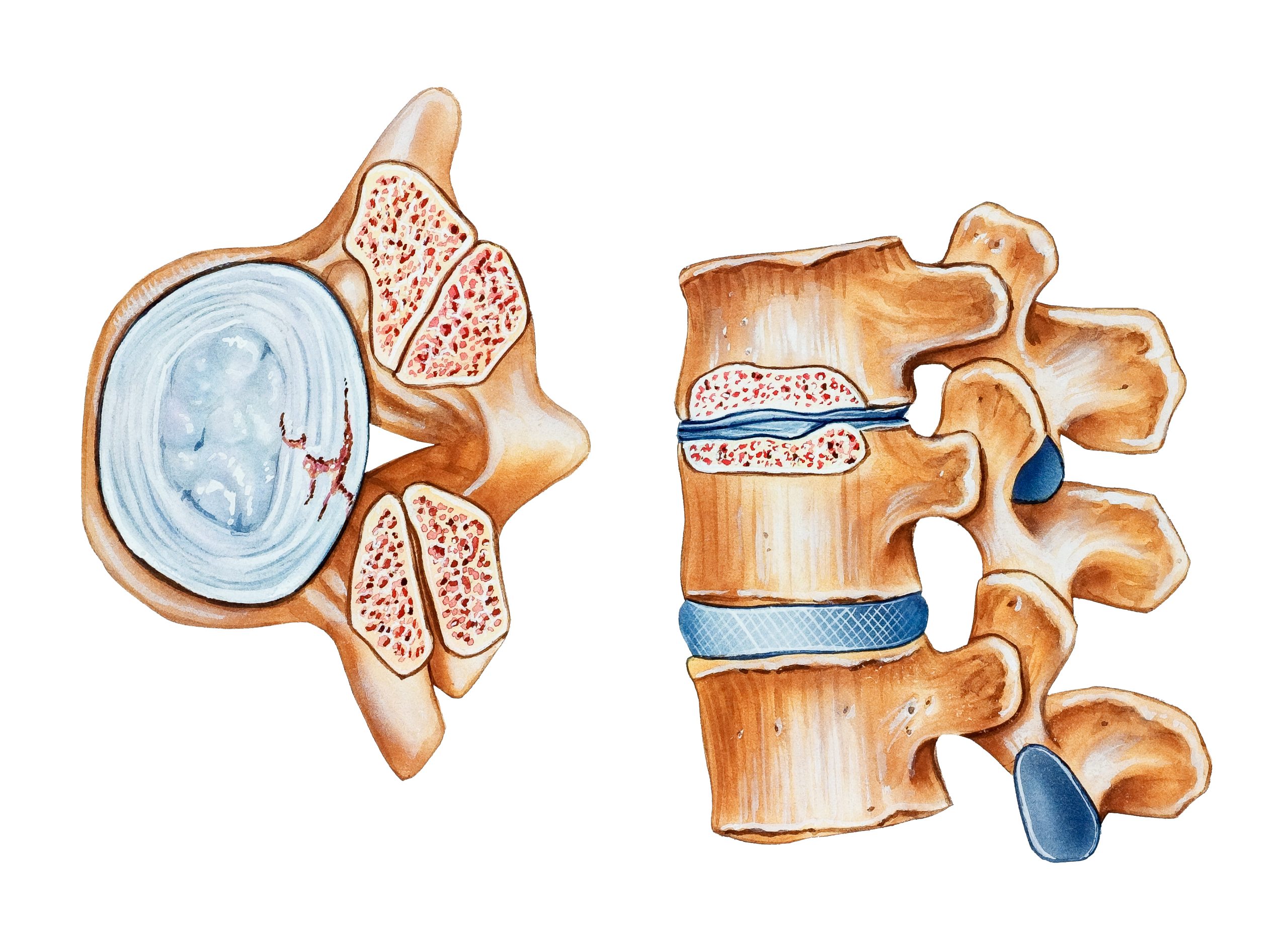
Lumbar laminectomy is a widely performed spinal surgery aimed at relieving pressure on the spinal cord or nerves in the lower back (lumbar spine). This procedure is particularly effective for treating conditions such as spinal stenosis, where the spinal canal narrows, causing significant pain and discomfort. By removing part or all of the lamina—the back part of a vertebra—the spinal canal is widened, alleviating pressure on the nerves and improving overall function and mobility.
Procedure for Lumbar Laminectomy
Open Lumbar Laminectomy
Step-by-Step Process
-
Incision:
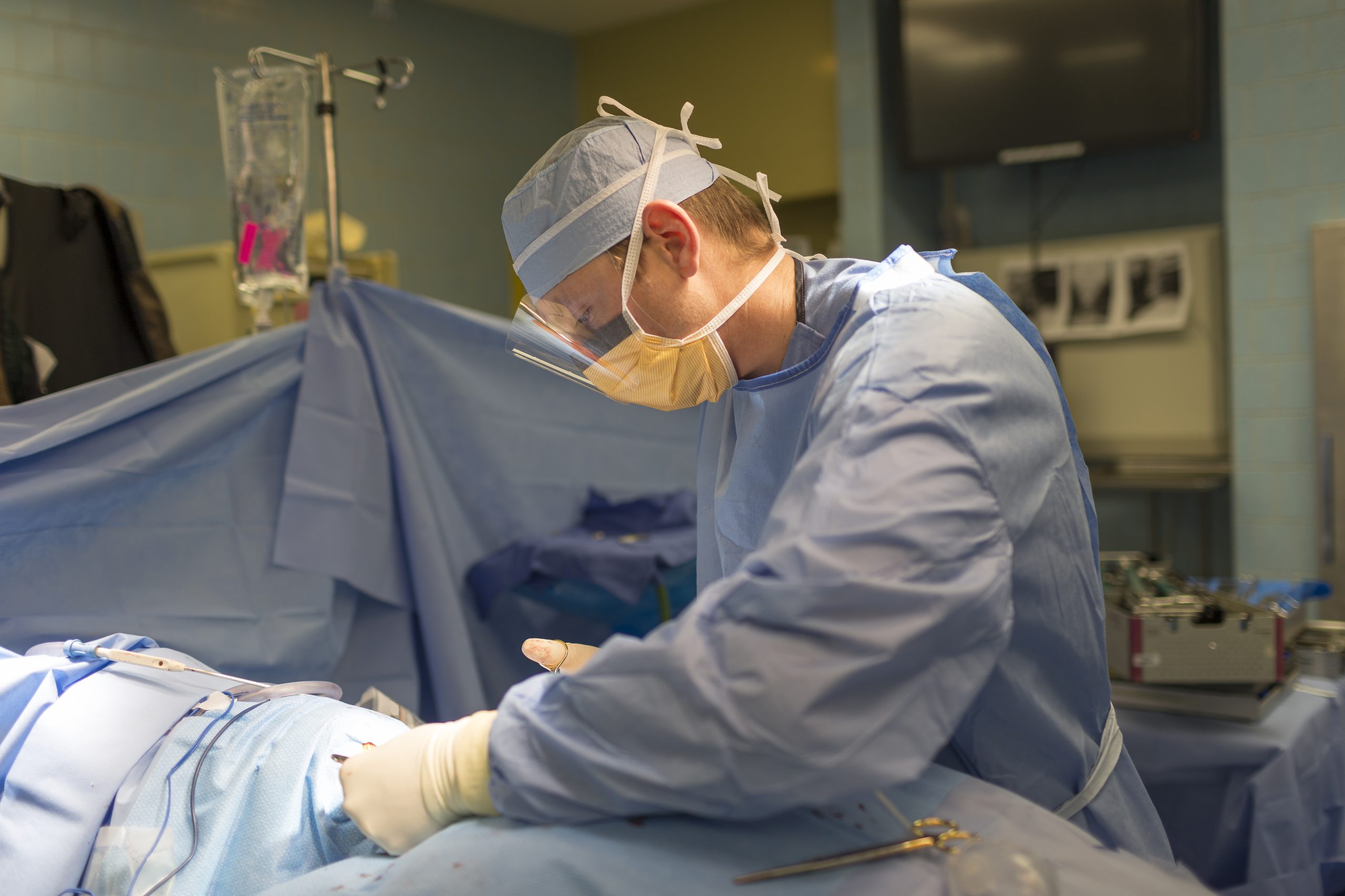
- The surgeon makes a vertical incision along the midline of the lower back. The length of the incision depends on the number of vertebrae being treated, typically ranging from 2 to 5 inches.
-
Muscle and Soft Tissue Retraction:
- The back muscles (erector spinae) and surrounding soft tissues are carefully moved aside using retractors. This process exposes the vertebrae and the lamina.
-
Removal of the Lamina:
- Using specialized surgical instruments such as bone rongeurs, high-speed burrs, and Kerrison rongeurs, the surgeon removes the lamina (the back part of the vertebra). This step widens the spinal canal and alleviates pressure on the spinal cord and nerve roots.
- In some cases, the spinous process (the bony projection off the back of each vertebra) may also be removed to provide additional space.
-
Decompression of the Spinal Canal:
- The surgeon ensures that the central canal, lateral recesses, and neural foramina (openings where nerve roots exit the spine) are adequately decompressed. This involves removing any thickened ligaments, bone spurs, or other structures compressing the nerves.
- Sometimes, a facetectomy (removal of part of the facet joint) or foraminotomy (widening of the foramen) may be performed to relieve nerve compression further.
-
Verification:
- The surgeon verifies that the spinal canal and nerve roots are adequately decompressed. This may involve using a surgical microscope or endoscope for better visualization.
-
Closure:
- Once decompression is complete, the retracted muscles and soft tissues are returned to their natural position. The incision is then closed with sutures or staples, and a sterile dressing is applied.
Recovery:
- Post-surgery, patients are monitored in the recovery room. Pain management is initiated, and patients are encouraged to walk soon after surgery. Most patients can go home the same day or after a short hospital stay, depending on the extent of the surgery and their overall health.
Minimally Invasive Lumbar Laminectomy
Step-by-Step Process
-
Small Incision:
- The surgeon makes a small incision, usually about 1 to 2 centimeters, over the affected vertebrae. This incision is significantly smaller than that used in open surgery, reducing tissue damage and scarring.
-
Tubular Retractor or Endoscope Insertion:
- A tubular retractor or endoscope is inserted through the small incision. The retractor gently spreads the muscles and soft tissues apart, creating a narrow tunnel to the spine. This approach minimizes muscle disruption and damage.
- The endoscope, equipped with a camera, provides the surgeon with a clear, magnified view of the surgical area on a monitor.
-
Removal of the Lamina:
- Specialized micro-surgical instruments are used to remove the lamina through the narrow tube. The use of a high-speed drill or burr may assist in precisely removing the bone.
- The surgeon may also remove any thickened ligaments, bone spurs, or other structures compressing the nerves.
-
Decompression of the Spinal Canal:
- Similar to the open technique, the goal is to decompress the spinal canal and nerve roots. This involves ensuring that the central canal, lateral recesses, and neural foramina are adequately widened.
- The endoscopic view allows for precise navigation and targeted removal of the compressive structures.
-
Verification:
- The surgeon verifies the decompression through the endoscope, ensuring all necessary structures are relieved of pressure. Real-time imaging, such as fluoroscopy, may be used to guide the procedure and confirm the correct levels.
-
Closure:
- The tubular retractor or endoscope is carefully removed. The small incision is closed with sutures or surgical glue, and a sterile dressing is applied.
Recovery:
- Minimally invasive procedures typically allow for a faster recovery. Patients experience less postoperative pain and shorter hospital stays, often being discharged the same day.
- Physical therapy may begin sooner compared to open surgery, aiding in a quicker return to normal activities.
Comparison of Open vs. Minimally Invasive Lumbar Laminectomy
- Incision Size: Open surgery involves a larger incision (2-5 inches) versus a small incision (1-2 cm) in minimally invasive surgery.
- Muscle Disruption: Open surgery requires more extensive muscle retraction, while minimally invasive techniques use a tubular retractor to minimize muscle damage.
- Visualization: Both methods aim to decompress the spinal canal, but minimally invasive surgery uses an endoscope for a magnified view, allowing for precise bone and tissue removal.
- Recovery: Minimally invasive surgery often results in less postoperative pain, shorter hospital stays, and quicker recovery times.
By offering detailed insight into both open and minimally invasive lumbar laminectomy procedures, Synergy Health Partners ensures that patients are well-informed about their surgical options and can make educated decisions about their spinal health.
Who is a Good Candidate for Lumbar Laminectomy?
- Persistent Symptoms: Patients with chronic back pain, leg pain, or weakness due to lumbar spinal stenosis that has not responded to conservative treatments like physical therapy, medications, or injections.
- Neurological Decline: Individuals showing signs of progressive neurological deficits, such as worsening leg strength or sensation.
- Spinal Instability: Those with spinal instability who might also require spinal fusion to prevent further complications.
- Thorough Medical Evaluation: Candidates undergo a detailed medical evaluation, including imaging studies and physical exams, to determine if lumbar laminectomy is the most suitable option.


Success and Risks
- Success Rate: Lumbar laminectomy generally has a high success rate, providing significant and lasting relief from symptoms of spinal stenosis.
- Risks and Complications: Like all surgeries, lumbar laminectomy carries some risks, including infection, bleeding, and nerve damage. These risks are typically low and manageable with proper care and monitoring.
Conditions Treated by Lumbar Laminectomy
Spine Surgeons That Offer Lumbar Laminectomy
Find a Location
MENDELSON KORNBLUM PAIN MANAGEMENT - LIVONIA
For appointments contact
Scheduling: 855.750.5757
For billing questions
Billing: 586.439.6242
Fax: 734.542.0220


